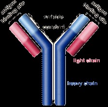
Nanone interactions in antibody of living systems
Abstract
One of the facts about how nanoparticle assemble and act is revealed using carbon value in biomolecule of living system here. This is how the biomolecules interact to bring about a micro or even macro level interaction in system of interest. This study shows micro level understanding can be better utilized from carbon analysis at nano level. I plan to extend this phenomena of change from nano to micro for building large scale applications in human nature. Applications include corrections in both at sequence and structure level for permanent recovery of defective one, adding flavor to the existing biomolecule for faster delivery or recovery etc. I have demonstrated here the active role played by carbon and all. This might be extended to another system of setup where new applications yet to be created. One can extend this phenomena of change from nano to large scale one.
References
[2] Indupriya R, Meenal R, and Rajasekaran E, Existence of carbon domain alters bond orders in protein, Int J Inno Eng Tech, 2019; 13: 128-32.
[3] Rajasekaran E, Domains based in carbon dictate here the possible arrangement of all chemistry for biology, Int. J. Mol Biol-Open Access, 2018; 3: 240-43.
[4] Rajasekaran E, Meenal R, and Indupriya R Study on aquaporin proves to be the carbon in protein-protein interface playing in tetramerisation, High Tech Lett, 2020; 26(5), 292-98.
[5] Rajasekaran E, Meenal R, and Indupriya R Carbon role in the form of action of intelligence in the living being, J Study Res, 2020; 12: 66-72.
[6] Rajasekaran E, Meenal R, Michael PA, and Indupriya R Existence of nano level force in protein plays applications of maximum untold understanding of life form, Int J Eng Adv Tech, 2019; 9: 3722-26.
[7] Rajasekaran E, Meenal R, Indupriya R, Prabakaran R, Boobalan S, Jayato N, Sivakumar K, Kalaivani T, Rathika GM, Saranya K, and Brindha G Existence of cohesive force explains all phenomena that are in material which holds strong bond of all forces of attraction: A case study with carbon material, AIP Conference Proceedings, 2019; 2087, 020015.
[8] Rajasekaran E, Indupriya R, and Meenal R, Domain formation in regions of protein probe interaction, Int J Mol Biol Open Access, 2019; 4: 167‒69.
[9] Rajasekaran E, Vinobha CS, Vijayasarathy M, Senthil R and Sankarganesh P, The nature of proteins, Int asso comput sci and info tech, (IACSIT-SC), Singapore, 2009; 464-65.
[10] Rajasekaran E, Scale for nature of hydrophobic interactions in proteins, J Proteomics and Bioinform, 2013; 6(7), 31.
[11] Rajasekaran E, CARd: Carbon distribution analysis program for protein sequences, Bioinformation, 2012; 8(11), 508-12.
[12] Rajasekaran E, Akila K, Vijayasarathy M, Vinobha CS, Senthil R and Anandagopu P, CARd-3D: Carbon distribution in 3D structure program for globular proteins, Bioinformation, 2014; 10(3), 138-43.
[13] Rajasekaran E, Kavitha V, Ganeshbabu P, Prabakaran R, Meenal R, and Indupriya R, Nature of amino acid sequence instruct carbon value to be adopted in protein 3D structure. IEEE Access, 2019; 1054-60.
[14] Rajasekaran E and Indupriya R, Who power sickle cell disease: Carbon domain analysis tells all because of design in protein 3D arbitrary internal carbon domain (COD) arrangement, Int J Mol Biol Open Access, 2019; 4: 85‒88.
[15] Rajasekaran E, John, SN and Vennila J, Carbon distribution in protein local structure direct superoxide dismutase to disease way, J Proteins and Proteomics, 2012; 3(2), 99-104.
[16] Rajasekaran E, Akila K and Vijayasarathy M, Allotment of carbon is responsible for disorders in proteins, Bioinformation, 2011; 6(8), 291-92.
[17] Amri E, Mamboya AF, Nsimama PD and Rajasekaran E, Role of carbon in crystal structures of wild-type and mutated form of dihydrofolate reductase-thymidylate synthase of P. falciparum, Int J App Bio and Pharma Tech, 2012; 3(3), 1-6.
[18] John SN, Anisha J, Kaarthik AN, Vennila, J and Rajasekaran E, Carbon and protein’s half-life, Int J Bioinfo, 2011; 4(2), 23-24.
[19] Nsimama PD, Mamboya AF, Amri E and Rajasekaran E, Correlation between the mutated color tunings and carbon distributions in luciferase bioluminescence, J Comput Intelli in Bioinfo, 2012; 5(2), 105-12.
[20] Rajasekaran E, Jency S and Panneerselvam K, Carbon profile of commercially important sericin protein of silkworm, Bombyx mori, J Adv Bioinfo Appln and Res, 2011; 2(3), 173-76.
[21] Rajasekaran E, Meenal R and Indupriya R, Paradigms in computer vision: Biology based carbon domain postulates nano electronic devices for generation next, In: Hemanth D., Smys S. (eds) Computational vision and bio inspired computing. Lecture notes in computational vision and biomechanics, 2019; 28. Springer, Cham.
[22] Mitchell LS, and Colwell LJ, Comparative analysis of nanobody sequence and structure data, Proteins: Structure, function and bioinformatics, 2018; 25497.


This work is licensed under a Creative Commons Attribution 4.0 International License.
Copyright for this article is retained by the author(s), with first publication rights granted to the journal.
This is an open-access article distributed under the terms and conditions of the Creative Commons Attribution license (http://creativecommons.org/licenses/by/4.0/).


1.png)


