Application of Dose Area Product (DAP) to Estimate Entrance Surface Dose (ESD) in Pediatric Chest X-Rays
ESD in Pediatric Chest X-rays
Abstract
Introduction: Given the high radiation tissue sensitivity of pediatric patients, it is necessary to monitor their received dose to optimize radiation protection. The first aim of this study was to evaluate the entrance surface dose (ESD) in pediatric patients undergoing a chest X-ray at the main hospital of Dezful, Iran. The second aim was to compare our results with the established dose reference levels (DRLs).
Materials and Methods: The studied population included 204 pediatric patients less than 15 years who were referred to as chest X-ray. A calibrated dose area product meter (DAP-meter) with permanent installation on the X-ray unit was used to radiation dose measurements. For each patient, the demographic data, exposure parameters and the dose read by DAP-meter were recorded and ESD was calculated using a standard mathematical formula.
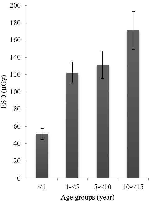
Results: The average value of ESD was 119 μGy in patients less than 15 years. This value was 51.3, 122.3, 131.5 and 171.2 μGy for the age groups for less than 1 year, 1 to 5 years, 5 to 10 years and 10 to 15 years, respectively. A statistically significant difference was seen between ESD values in different age groups (P<0.001), whereas no statistical difference was seen between ESD values in girls and boys (P =0.993).
Conclusion: Pediatric patients in hospital investigated (except age group less than 1 year) are subjected to unnecessary radiation exposure, especially due to the use of non-optimize X-ray protocols.
References
[2] Curtis JR. (2010). Computed tomography shielding methods: A literature review. Radiologic Technology, 81(5), 428-36.
[3] Hohl, C., Wildberger, J., Süß, C., Thomas, C., Mühlenbruch, G., Schmidt, T., et al. (2006). Radiation dose reduction to breast and thyroid during MDCT: effectiveness of an in-plane bismuth shield. Acta Radiologica, 47(6), 562-7. https://doi.org/10.1080/02841850600702150
[4] Kostova-Lefterova, D., Taseva, D., Hristova-Popova, J., & Vassileva, J. (2015). Optimisation of paediatric chest radiography. Radiation Protection Dosimetry, 165(1-4), 231-4. https://doi.org/10.1093/rpd/ncv119
[5] Toossi, M. T. B., & Malekzadeh, M. (2012). Radiation dose to newborns in neonatal intensive care units. Iranian Journal of Radiology, 9(3), 145-9. https://doi.org/10.5812/iranjradiol.8065
[6] Atalabi, O. M., BidemiI, A., Adekanmi, A. J., & Samuel, O. A. (2013). Entrance surface dose from pediatric diagnostic x-ray examinations in a developing world setting: are we'ALARA principle'compliant? British Journal of Medicine and Medical Research, 3(4), 2288-98. https://doi.org/10.9734/BJMMR/2013/4119
[7] Kim, B. H., Do, K. H., Goo, H. W., Yang, D. H., Oh, S. Y., Kim, H. J., et al. (2012). National survey of radiation doses of pediatric chest radiography in Korea: analysis of the factors affecting radiation doses. Korean Journal of Radiology, 13(5), 610-7. https://doi.org/10.3348/kjr.2012.13.5.610
[8] Nikupaavo, U., Kaasalainen, T., Reijonen, V., Ahonen, S. M., & Kortesniemi, M. (2015). Lens dose in routine head CT: comparison of different optimization methods with anthropomorphic phantoms. American Journal of Roentgenology, 204(1), 117-23. https://doi.org/10.2214/AJR.14.12763
[9] Freitas, M. B., & Yoshimura, E. M. (2009). Diagnostic reference levels for the most frequent radiological examinations carried out in Brazil. Revista Panamericana de Salud Pública, 25(2), 95-104. https://doi.org/10.1590/S1020-49892009000200001
[10] Jornet, N., Muñoz, J., Martin-Viera, J., Jurado, D., Pallerol, R., Gultresa, J., et al. Determination of entrance surface dose in standard explorations in radiodiagnostic. Retrieved January 2019, from https://pdfs.semanticscholar.org/f406/79f5061c9bf368feb0768bf68f3dce621f2d.pdf
[11] Ofori, K., Gordon, S. W., Akrobortu, E., Ampene, A. A., & Darko, E. O. (2014). Estimation of adult patient doses for selected X-ray diagnostic examinations. Journal of Radiation Research and Applied Sciences, 7(4), 459-62. https://doi.org/10.1016/j.jrras.2014.08.003
[12] Zewdu, M., Kadir, E., & Berhane, M. (2017). Assessment of pediatrics radiation dose from routine x-ray examination at Jimma University Hospital, Southwest Ethiopia. Ethiopian Journal of Health Sciences, 27(5), 481-90. https://doi.org/10.4314/ejhs.v27i5.6
[13] Gholami, M., Maziar, A., Khosravi, H., Ebrahimzadeh, F., & Mayahi, S. (2015). Diagnostic reference levels (DRLs) for routine X-ray examinations in Lorestan province, Iran. International Journal of Radiation Research, 13(1), 85-90.
[14] Faghihi, R., Mehdizadeh, S., Sina, S., Alizadeh, F. N., Zeinali, B., Kamyab, G. R., et al. (2011). Radiation dose to neonates undergoing X-ray imaging in special care baby units in Iran. Radiation protection dosimetry. 150(1), 55-9. https://doi.org/10.1093/rpd/ncr373
[15] Ademola, A., Obed, R., Adejumobi, C., Abodunrin, O., Alabi, O., & Oladapo, M. (2013). Assessment of Entrance Skin Dose in routine x-ray examinations of chest, skull, abdomen and pelvis of children in five selected hospitals in Nigeria. IOSR Journal of Applied Physics. 5(2), 47-50. https://doi.org/10.9790/4861-0524750
[16] Kisielewicz, K., Truszkiewicz, A., Wach, S., & Wasilewska–Radwańska, M. (2011). Evaluation of dose area product vs. patient dose in diagnostic X-ray units. Physica Medica, 27(2), 117-20. https://doi.org/10.1016/j.ejmp.2010.07.001
[17] Hart, D., Jones, D., & Wall B. (1994). Estimation of effective dose in diagnostic radiology from entrance surface dose and dose-area product measurements. National Radiological Protection Board, 71(849), 994-5. https://doi.org/10.1259/bjr.71.849.10195022
[18] Hart, D., Jones, D., & Wall B. (1996). Coefficients for estimating effective doses from paediatric x-ray examinations: National Radiological Protection Board Oxon.
[19] Martin, C. (1995). Measurement of patient entrance surface dose rates for fluoroscopic x-ray units. Physics in Medicine & Biology, 40(5), 823-34. https://doi.org/10.1088/0031-9155/40/5/008
[20] McParland, B. (1998). Entrance skin dose estimates derived from dose-area product measurements in interventional radiological procedures. The British Journal of Radiology, 71(852), 1288-95. https://doi.org/10.1259/bjr.71.852.10319003
[21] Smans K, Struelens L, Smet M, Bosmans H, Vanhavere F. (2008). Patient dose in neonatal units. Radiation Protection Dosimetry, 131(1), 143-7. https://doi.org/10.1093/rpd/ncn237
[22] European Union. European Commission. (1996). Directorate-General XII-Science R, Development. European guidelines on quality criteria for diagnostic radiographic images in paediatrics: Office for Official Publications of the European Communities.

This work is licensed under a Creative Commons Attribution 4.0 International License.
Copyright for this article is retained by the author(s), with first publication rights granted to the journal.
This is an open-access article distributed under the terms and conditions of the Creative Commons Attribution license (http://creativecommons.org/licenses/by/4.0/).


1.png)