Advances in the Pathogenesis and Treatment of Esophageal Cancer with Traditional Chinese Medicine
Abstract
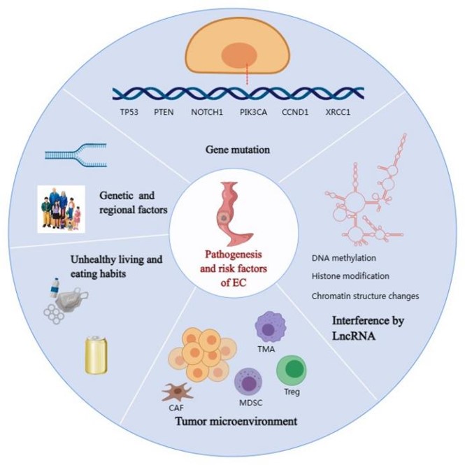
Esophageal cancer (EC) is one of the common malignancies with high morbidity and mortality. Despite the effectiveness of modern medical interventions such as surgery, radiotherapy, and chemotherapy in treating EC, complications and adverse reactions still exist. Traditional Chinese medicine (TCM) plays a crucial role in preventing and treating malignant tumors by employing its unique approach based on syndrome differentiation and the concept of wholism. Numerous fundamental experiments and clinical studies have provided substantial evidence supporting the beneficial effects of TCM in managing EC. The pathogenesis of EC and the potential mechanism of TCM intervention in EC treatment are summarized in this review, aiming to offer a novel perspective for EC therapy.
References
[2] Feng, R. M., Zong, Y. N., Cao, S. M., Xu, R. H., & Zheng, R. S. (2019). Current cancer situation in China: Good or bad news from the 2018 Global Cancer Statistics. Cancer Communications, 39(1), 22–31. https://doi.org/10.1186/s40880-019-0368-6
[3] Hu, Y., Correa, A. M., Hoque, A., Guan, B., Liang, W., Jian, W., & Li, R. (2011). Prognostic significance of differentially expressed miRNAs in esophageal cancer. International Journal of Cancer, 128(1), 132–143. https://doi.org/10.1002/ijc.25330
[4] Liu, J. B., & Yue, J. Y. (2014). Preliminary study on the mechanism of oridonin-induced apoptosis in human squamous cell oesophageal carcinoma cell line EC9706. Journal of International Medical Research, 42(4), 984–992. https://doi.org/10.1177/0300060513507389
[5] Wu, T., Yang, X., Zeng, X., & Liu, J. (2009). Traditional Chinese medicinal herbs in the treatment of patients with esophageal cancer: A systematic review. Gastroenterology Clinics of North America, 38(1), 153–167. https://doi.org/10.1016/j.gtc.2009.01.006
[6] Sarkar, F. H., Li, Y. W., Wang, Z. W., & Kong, J. S. (2010). Lesson learned from nature for the development of novel anti-cancer agents: Implication of isoflavone, curcumin, and their synthetic analogs. Current Pharmaceutical Design, 16(16), 1801–1812. https://doi.org/10.2174/138161210791208956
[7] Uhlenhopp, D. J., Then, E. O., Sunkara, T., & Reddy, D. N. (2020). Epidemiology of esophageal cancer: Update in global trends, etiology and risk factors. Clinical Journal of Gastroenterology, 13(6), 1010–1021. https://doi.org/10.1007/s12328-020-01237-x
[8] Conroy, M. J., Kennedy, S. A., Doyle, S. L., & Mulcahy, H. (2021). A study of the immune infiltrate and patient outcomes in esophageal cancer. Carcinogenesis, 42(3), 395–404. https://doi.org/10.1093/carcin/bgaa101
[9] Greenblatt, M. S., Bennett, W. P., Hollstein, M., & Harris, C. C. (1994). Mutations in the p53 tumor suppressor gene: Clues to cancer etiology and molecular pathogenesis. Cancer Research, 54(18), 4855–4878.
[10] Reid, B. J., Prevo, L. J., Galipeau, P. C., & Mark, J. (2001). Predictors of progression in Barrett's esophagus II: Baseline 17p (p53) loss of heterozygosity identifies a patient subset at increased risk for neoplastic progression. American Journal of Gastroenterology, 96(10), 2839–2848. https://doi.org/10.1016/S0002-9270(01)02798-8
[11] Gamieldien, W., Victor, T. C., Mugwanya, D., & van der Merwe, J. M. (1998). p53 and p16/CDKN2 gene mutations in esophageal tumors from a high-incidence area in South Africa. International Journal of Cancer, 78(5), 544–549. https://doi.org/10.1002/(SICI)1097-0215(19981123)78:5<544::AID-IJC3>3.0.CO;2-T
[12] Robert, V., Michel, P., Flaman, J. M., & Frebourg, T. (2000). High frequency in esophageal cancers of p53 alterations inactivating the regulation of genes involved in cell cycle and apoptosis. Carcinogenesis, 21(4), 563–565. https://doi.org/10.1093/carcin/21.4.563
[13] Oki, E., Zhao, Y., Yoshida, R., & Saeki, H. (2009). The difference in p53 mutations between cancers of the upper and lower gastrointestinal tract. Digestion, 79(Suppl 1), 33–39. https://doi.org/10.1159/000167864
[14] Smith, U. (2012). PTEN-linking metabolism, cell growth, and cancer. New England Journal of Medicine, 367(11), 1061–1063. https://doi.org/10.1056/NEJMe1208934
[15] Qu, W., Fu, J. D., Yang, F., & Yang, B. (2015). Clinical implications of PTEN and VEGF expression status, as well as microvessel density in esophageal squamous cell carcinoma. Oncology Letters, 10(3), 1409–1415. https://doi.org/10.3892/ol.2015.3431
[16] Chang, D., Wang, T. Y., Li, H. C., & Fu, Y. (2007). Prognostic significance of PTEN expression in esophageal squamous cell carcinoma from Linzhou City, a high incidence area of northern China. Diseases of the Esophagus, 20(6), 491–496. https://doi.org/10.1111/j.1442-2050.2007.00695.x
[17] Tachibana, M., Shibakita, S., Ohno, S., & Tanamura, H. (2002). Expression and prognostic significance of PTEN product protein in patients with esophageal squamous cell carcinoma. Cancer, 94(7), 1955–1960. https://doi.org/10.1002/cncr.0678
[18] Zhou, Y., Zhang, T., Zhao, J. B., & Wang, W. (2010). The adenovirus-mediated transfer of PTEN inhibits the growth of esophageal cancer cells in vitro and in vivo. Bioscience, Biotechnology, and Biochemistry, 74(4), 736–740. https://doi.org/10.1271/bbb.90787
[19] Sun, Z., Ji, N., Bi, M. M., & Wang, Q. (2015). Negative expression of PTEN identifies high risk for lymphatic-related metastasis in human esophageal squamous cell carcinoma. Oncology Reports, 33(6), 3024–3032. https://doi.org/10.3892/or.2015.3928
[20] Katoh, M. (2007). Notch signaling in gastrointestinal tract. International Journal of Oncology, 30(1), 247–251. https://doi.org/10.3892/ijo.30.1.247
[21] Zhang, K. J., Lu, Q. Y., Niu, X. Q., & Tang, G. G. (2009). Notch1 signaling inhibits growth of EC109 esophageal carcinoma cells through downmodulation of HPV18 E6/E7 gene expression. Acta Pharmacologica Sinica, 30(2), 153–158. https://doi.org/10.1038/aps.2008.16
[22] Song, B., Cui, H. Y., Li, Y. P., Wang, Z., & Chen, X. (2016). Mutually exclusive mutations in NOTCH1 and PIK3CA associated with clinical prognosis and chemotherapy responses of esophageal squamous cell carcinoma in China. Oncotarget, 7(3), 3599–3613. https://doi.org/10.18632/oncotarget.6120
[23] Samuels, Y., & Ericson, K. (2006). Oncogenic PI3K and its role in cancer. Current Opinion in Oncology, 18(1), 77–82. https://doi.org/10.1097/01.cco.0000198021.99347.b9
[24] Shigaki, H., Baba, Y., Watanabe, M., & Nomura, M. (2013). PIK3CA mutation is associated with a favorable prognosis among patients with curatively resected esophageal squamous cell carcinoma. Clinical Cancer Research, 19(9), 2451–2459. https://doi.org/10.1158/1078-0432.CCR-12-3559
[25] Hou, J., Jiang, D. X., Zhang, J., & Guo, W. P. (2014). Frequency, characterization, and prognostic analysis of PIK3CA gene mutations in Chinese esophageal squamous cell carcinoma. Human Pathology, 45(2), 352–358. https://doi.org/10.1016/j.humpath.2013.09.011
[26] Akagi, M., Miyashita, H., Makino, H., & Nishino, M. (2009). Overexpression of PIK3CA is associated with lymph node metastasis in esophageal squamous cell carcinoma. International Journal of Oncology, 34(3), 767–775. https://doi.org/10.3892/ijo_00000202
[27] Komatsu, S., Ichikawa, D., Hirajima, S., & Okamoto, K. (2014). Clinical impact of predicting CCND1 amplification using plasma DNA in superficial esophageal squamous cell carcinoma. Digestive Diseases and Sciences, 59(6), 1152–1159. https://doi.org/10.1007/s10620-013-3005-2
[28] Zhao, J., Li, L., Wei, S., & Li, R. (2012). Clinicopathological and prognostic role of cyclin D1 in esophageal squamous cell carcinoma: A meta-analysis. Diseases of the Esophagus, 25(6), 520–526. https://doi.org/10.1111/j.1442-2050.2011.01278.x
[29] Solomon, D. A., Wang, Y., Fox, S. R., & Chen, L. J. (2003). Cyclin D1 splice variants: Differential effects on localization, RB phosphorylation, and cellular transformation. Journal of Biological Chemistry, 278(32), 30339–30347. https://doi.org/10.1074/jbc.M303969200
[30] Li, S., Deng, Y., You, J. P., & Liu, X. (2013). XRCC1 Arg399Gln, Arg194Trp, and Arg280His polymorphisms in esophageal cancer risk: A meta-analysis. Digestive Diseases and Sciences, 58(7), 1880–1890. https://doi.org/10.1007/s10620-013-2569-1
[31] Wei, B., Han, Q., Xu, L. J., & Zheng, J. G. (2015). Effects of JWA, XRCC1 and BRCA1 mRNA expression on molecular staging for personalized therapy in patients with advanced esophageal squamous cell carcinoma. BMC Cancer, 15, 331. https://doi.org/10.1186/s12885-015-1364-0
[32] Chen, J. L., Lin, Z. X., Qin, Y. S., & Zhong, Y. H. (2019). Overexpression of long noncoding RNA LINC01419 in esophageal squamous cell carcinoma and its relation to the sensitivity to 5-fluorouracil by mediating GSTP1 methylation. Therapeutic Advances in Medical Oncology, 11, 1–17. https://doi.org/10.1177/1758835919838958
[33] Wu, Y., Hu, L., Lian, Y., & Mo, Y. (2017). Up-regulation of lncRNA CASC9 promotes esophageal squamous cell carcinoma growth by negatively regulating PDCD4 expression through EZH2. Molecular Cancer, 16(1), 150. https://doi.org/10.1186/s12943-017-0715-7
[34] Lian, Y., Chen, X., Wu, Y., & Mo, Y. (2018). LncRNA CASC9 promotes esophageal squamous cell carcinoma metastasis through upregulating LAMC2 expression by interacting with the CREB-binding protein. Cell Death & Differentiation, 25(11), 1980–1995. https://doi.org/10.1038/s41418-018-0084-9
[35] Jayaraman, P., Parikh, F., Lopez-Rivera, E., & Sharma, S. (2012). Tumor-expressed inducible nitric oxide synthase controls induction of functional myeloid-derived suppressor cells through modulation of vascular endothelial growth factor release. Journal of Immunology, 188(11), 5365–5376. https://doi.org/10.4049/jimmunol.1103553
[36] Li, P. P., Chen, X. F., Qin, G. H., & Jiang, Z. W. (2018). Maelstrom directs myeloid-derived suppressor cells to promote esophageal squamous cell carcinoma progression via activation of the Akt1/RelA/IL8 signaling pathway. Cancer Immunology Research, 6(10), 1246–1259. https://doi.org/10.1158/2326-6066.CIR-17-0415
[37] Wang, G. C., Liu, G. Y., Liu, Y., & Su, J. P. (2012). FOXP3 expression in esophageal cancer cells is associated with poor prognosis in esophageal cancer. Hepato-Gastroenterology, 59(119), 2186–2191. https://doi.org/10.5754/hge11961
[38] Nabeki, B., Ishigami, S., Uchikado, Y., & Kita, Y. (2015). Interleukin-32 expression and Treg infiltration in esophageal squamous cell carcinoma. Anticancer Research, 35(5), 2941–2947.
[39] Xia, M., Zhao, M. Q., Wu, K., & Zhang, W. (2013). Investigations on the clinical significance of FOXP3 protein expression in cervical oesophageal cancer and the number of FOXP3+tumour-infiltrating lymphocytes. Journal of International Medical Research, 41(4), 1002–1008. https://doi.org/10.1177/0300060513488504
[40] Xu, T. Y., Duan, Q. H., Wang, G., & Liu, Q. (2011). CD4+CD25(high) regulatory T cell numbers and FOXP3 mRNA expression in patients with advanced esophageal cancer before and after chemotherapy. Cell Biochemistry and Biophysics, 61(2), 389–392. https://doi.org/10.1007/s12013-011-9197-1
[41] Shitara, K., & Nishikawa, H. (2018). Regulatory T cells: A potential target in cancer immunotherapy. Annals of the New York Academy of Sciences, 1417(1), 104–115. https://doi.org/10.1111/nyas.13625
[42] Mitra, A. K., Zillhardt, M., Hua, Y., & Madi, A. (2012). MicroRNAs reprogram normal fibroblasts into cancer-associated fibroblasts in ovarian cancer. Cancer Discovery, 2(12), 1100–1108. https://doi.org/10.1158/2159-8290.CD-12-0206
[43] Marsh, T., Pietras, K., & McAllister, S. S. (2013). Fibroblasts as architects of cancer pathogenesis. Biochimica et Biophysica Acta (BBA)-Molecular Basis of Disease, 1832(7), 1070–1078. https://doi.org/10.1016/j.bbadis.2012.10.013
[44] Hiroki, S., Shuji, T., Yuto, S., & Shinsuke, E. (2021). Achalasia and esophageal cancer: A large database analysis in Japan. Journal of Gastroenterology, 56(4), 360–370. https://doi.org/10.1007/s00535-021-01763-6
[45] Mao, W. M., Zheng, W. H., & Ling, Z. Q. (2011). Epidemiologic risk factors for esophageal cancer development. Asian Pacific Journal of Cancer Prevention, 12(10), 2461–2466.
[46] Cho, J. H., Shin, C. M., Han, K. D., & Kim, J. H. (2020). Abdominal obesity increases risk for esophageal cancer: A nationwide population-based cohort study of South Korea. Journal of Gastroenterology, 55(3), 307–316. https://doi.org/10.1007/s00535-019-01648-9
[47] Cui, W., Wu, Y., Wei, S., & Hu, J. (2017). Inhibitory effects of p-hydroxylcinnamaldehyde on esophageal cancer xenograft tumor in vivo. Chinese Journal of Cancer Biotherapy, 24(2), 145–150.
[48] Wang, L. F., Lu, A., Meng, F., & Li, R. Y. (2012). Inhibitory effects of lupeal acetate of Cortex periplocae on N-nitrosomethylbenzylamine-induced rat esophageal tumorigenesis. Oncology Letters, 4(2), 231–236. https://doi.org/10.3892/ol.2012.717
[49] Liu, R. P., Wang, X. Q., Wang, J., & Wang, Y. (2022). Oroxin A reduces oxidative stress, apoptosis, and autophagy and improves the developmental competence of porcine embryos in vitro. Reproduction in Domestic Animals, 57(10), 1255–1266. https://doi.org/10.1111/rda.14200
[50] Shipa, V. S., Shams, R., Dash, K. K., & Islam, M. (2023). Phytochemical properties, extraction, and pharmacological benefits of Naringin: A review. Molecules, 28(15), 5623. https://doi.org/10.3390/molecules28155623
[51] Si, F. C. (2021). Effects of different ethanol elutefractions of Qigesan, Tongyoutang, Shashenmaidongtang and Buqiyunpitang on expression of secretory proteins in esophageal carcinoma EC9706 cell. Cancer Research, 80(16), 1856. https://doi.org/10.1158/1538-7445.AM2020-1856
[52] Wang, S., Gao, X., Li, J., & Zhang, Q. (2022). The anticancer effects of curcumin and clinical research progress on its effects on esophageal cancer. Frontiers in Pharmacology, 13, 1058070. https://doi.org/10.3389/fphar.2022.1058070
[53] Zhan, Y., Li, R., Feng, C. L., & Chen, Y. (2020). Chlorogenic acid inhibits esophageal squamous cell carcinoma growth in vitro and in vivo by downregulating the expression of BMI1 and SOX2. Biomedicine & Pharmacotherapy, 121, 109602. https://doi.org/10.1016/j.biopha.2019.109602
[54] Song, L., Xiong, P. Y., Zhang, W., & Li, J. (2022). Mechanism of Citri Reticulatae Pericarpium as an anticancer agent from the perspective of flavonoids: A review. Molecules, 27(17), 5622. https://doi.org/10.3390/molecules27175622
[55] Jurikova, T., Sochor, J., Rop, O., & Mlcek, J. (2012). Polyphenolic profile and biological activity of Chinese Hawthorn (Crataegus pinnatifida BUNGE) fruits. Molecules, 17(12), 14490–14509. https://doi.org/10.3390/molecules171214490
[56] Wu, D., Li, J., Hu, X., & Zhu, S. (2018). Hesperetin inhibits Eca-109 cell proliferation and invasion by suppressing the PI3K/AKT signaling pathway and synergistically enhances the anti-tumor effect of 5-fluorouracil on esophageal cancer in vitro and in vivo. RSC Advances, 8(43), 24434–24443. https://doi.org/10.1039/C8RA00956B
[57] Wang, H. Y., Hu, H. Y., Rong, H., & Liu, X. J. (2019). Effects of compound Kushen injection on pathology and angiogenesis of tumor tissues. Oncology Letters, 17(2), 2278–2282. https://doi.org/10.3892/ol.2018.9861
[58] Han, Z., Guo, J. M., Meng, F. B., & Zhang, Y. (2021). Genetic toxicology and safety pharmacological evaluation of forsythin. Evidence-Based Complementary and Alternative Medicine, 2021, 6610793. https://doi.org/10.1155/2021/6610793
[59] Chen, X. H., Wang, S. W., Zhang, L., & Li, R. Y. (2022). Celastrol inhibited human esophageal cancer by activating DR5-dependent extrinsic and Noxa/Bim-dependent intrinsic apoptosis. Frontiers in Pharmacology, 13, 873166. https://doi.org/10.3389/fphar.2022.873166
[60] Cai, Y., Gao, K. W., Peng, B., & Wang, J. H. (2021). Alantolactone: A natural plant extract as a potential therapeutic agent for cancer. Frontiers in Pharmacology, 12, 781033. https://doi.org/10.3389/fphar.2021.781033
[61] Pu, Y., Jin, P., Liu, L., & Li, Y. (2021). Dysosma versipellis extract inhibits esophageal cancer progression through the Wnt signaling pathway. Evidence-Based Complementary and Alternative Medicine, 2021, 1221899. https://doi.org/10.1155/2021/1221899
[62] Yu, J., Wang, W., Liu, B., & Li, Z. (2021). Demethylzelasteral inhibits proliferation and EMT via repressing Wnt/β-catenin signaling in esophageal squamous cell carcinoma. Journal of Cancer, 12(13), 3967–3975. https://doi.org/10.7150/jca.45493
[63] Ju, Q. Q., Shi, Q. Y., Liu, C., & Wei, B. (2023). Bufalin suppresses esophageal squamous cell carcinoma progression by activating the PIAS3/STAT3 signaling pathway. Journal of Thoracic Disease, 15(4), 2141–2160. https://doi.org/10.21037/jtd-23-486
[64] Chen, W. X., Lu, Y., Chen, G. Y., & Li, S. (2013). Molecular evidence of cryptotanshinone for treatment and prevention of human cancer. Anti-Cancer Agents in Medicinal Chemistry, 13(7), 979–987. https://doi.org/10.2174/18715206113139990115
[65] Wang, P. L., Yang, J., Zhu, Z. F., & Wu, C. Y. (2018). Marsdenia tenacissima: A review of traditional uses, phytochemistry and pharmacology. American Journal of Chinese Medicine, 46(7), 1449–1480. https://doi.org/10.1142/S0192415X18500751
[66] Li, Q. R., Ma, Q., Xu, L., & Wei, B. (2021). Human telomerase reverse transcriptase as a therapeutic target of dihydroartemisinin for esophageal squamous cancer. Frontiers in Pharmacology, 12, 769787. https://doi.org/10.3389/fphar.2021.769787
[67] Han, S. C., Gou, Y. J., Jin, D. C., & Lee, D. W. (2018). Effects of icaritin on the physiological activities of esophageal cancer stem cells. Biochemical and Biophysical Research Communications, 504(4), 792–796. https://doi.org/10.1016/j.bbrc.2018.08.060
[68] Han, Y. C., Fan, X. Q., Fan, L. Y., & He, J. (2023). Liujunzi decoction exerts potent antitumor activity in oesophageal squamous cell carcinoma by inhibiting miR-34a/STAT3/IL-6R feedback loop, and modifies antitumor immunity. Phytomedicine, 111, 154672. https://doi.org/10.1016/j.phymed.2023.154672
[69] Shen, Z., Xu, L., Li, J., & Zhang, J. (2017). Capilliposide C sensitizes esophageal squamous carcinoma cells to oxaliplatin by inducing apoptosis through the PI3K/Akt/mTOR pathway. Medical Science Monitor, 23, 2096–2103. https://doi.org/10.12659/MSM.902704
[70] Wang, T., Wang, J., Ren, W., & Yang, Y. (2020). Combination treatment with artemisinin and oxaliplatin inhibits tumorigenesis in esophageal cancer EC109 cell through Wnt/β-catenin signaling pathway. Thoracic Cancer, 11(8), 2316–2324. https://doi.org/10.1111/1759-7714.13570
[71] Ying, J., Zhang, M., Qiu, X., & Li, Q. (2018). The potential of herb medicines in the treatment of esophageal cancer. Biomedicine & Pharmacotherapy, 103, 381–390. https://doi.org/10.1016/j.biopha.2018.04.088
[72] Cusack, J. C., Liu, R., Houston, M., & Meng, F. (2001). Enhanced chemosensitivity to CPT-11 with proteasome inhibitor PS-341: Implications for systemic nuclear factor-kappa B inhibition. Cancer Research, 61(9), 3535–3540.
[73] Voboril, R., Hochwald, S. N., Li, J., & Wang, J. (2004). Inhibition of NF-kappa B augments sensitivity to 5-fluorouracil/folinic acid in colon cancer. Journal of Surgical Research, 120(2), 178–188. https://doi.org/10.1016/j.jss.2003.11.023
[74] Zhang, D., Ni, M. W., Wu, J. R., & Li, Y. (2019). The optimal Chinese herbal injections for use with radiotherapy to treat esophageal cancer: A systematic review and Bayesian network meta-analysis. Frontiers in Pharmacology, 9, 1470. https://doi.org/10.3389/fphar.2018.01470
[75] Yang, X., Yang, B. X., Cai, J., & Meng, F. (2013). Berberine enhances radiosensitivity of esophageal squamous cancer by targeting HIF-1 alpha in vitro and in vivo. Cancer Biology & Therapy, 14(11), 1068–1073. https://doi.org/10.4161/cbt.26426


This work is licensed under a Creative Commons Attribution 4.0 International License.
Copyright for this article is retained by the author(s), with first publication rights granted to the journal.
This is an open-access article distributed under the terms and conditions of the Creative Commons Attribution license (http://creativecommons.org/licenses/by/4.0/).


1.png)


