A Case Report of Chronic Kidney Disease Stage 5 Complicated with Uremic Tumor Calcinosis
Abstract
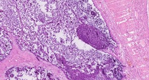
Uremic tumor calcinosis (UTC) represents an extremely rare complication associated with chronic kidney disease (CKD), prominently characterized by the occurrence of ectopic calcification. The incidence of UTC among uremic peritoneal dialysis patients has been estimated to be approximately 1.60%. This specific case report aims to illuminate the intricate diagnostic and therapeutic challenges encountered in a dialysis patient. A 74-year-old male, diagnosed with stage 5 CKD and undergoing peritoneal dialysis, presented with a considerable 12x5 cm soft tissue mass on his back. Through a comprehensive array of imaging studies and histopathological examinations, the mass was subsequently identified as UTC. The diagnostic process revealed elevated levels of Parathyroid hormone (PTH) at 445.100 pg/ml and phosphorus at 2.77mg/dl. The diagnosis was ultimately confirmed via surgical resection. The patient's condition was managed and stabilized through the administration of phosphate binders and calcitriol. This case significantly underlines the critical necessity of vigilant and continuous monitoring of calcium-phosphorus metabolism in patients with CKD. Furthermore, it highlights UTC as an important differential diagnosis consideration when confronted with soft tissue masses.
References
[2] Li, Y. P., Xu, H. Y., Liu, F. L., et al. (2018). Effect of parathyroidectomy on SHPT and tumor-like calcinosis and serum FGF23 levels in hemodialysis patients at the end of the treatment period. Journal of Difficult Diseases, 17(11), 1229–1232.
[3] Yano, H., & Kinjo, M. (2021). Turkish calcinosis. Cleveland Clinic Journal of Medicine, 88(4), 208–209.
[4] Yi, M. H., & Lu, J. (2022). A case of rare uremic tumor-like calcification syndrome. JPMI, 23(4), 431–432.
[5] Fatehi, M., Ahuja, C. S., Wang, S., et al. (2016). Uremic tumoral calcinosis in the cervical spine: Case report. Journal of Neurosurgery: Spine, 25(1), 26–30. https://doi.org/10.3171/2015.12.SPINE151085
[6] Xu, B., Luo, Y. C., Wang, S. B., et al. (2011). A case report of lumbar tumor-like calcinosis. Chinese Journal of Orthopedics, 19(21), 1842–1843.
[7] Jin, Q. J., Zhou, H. Y., & Lu, H. (2021). Intramuscular tumoral calcinosis near the arteriovenous fistula mimicking acute infection. Science Progress, 104(2), 1–10. https://doi.org/10.1177/00368504211018560
[8] Rosen, R. J., Fernandez, H. E., Shirazian, S., et al. (2021). Ultrasound findings of calciphylaxis. Kidney International, 100, 1144.
[9] Zheng, S. W., Sun, B., & Liu, C. (2019). Surgical treatment of uremic tumor-like calcinosis: A case report. Bone Setting of Traditional Chinese Medicine, 31(9), 74–77.
[10] Tian, F., Xing, G. Q., & Wang, Y. R. (2019). A case of giant tumor-like calcinosis in a long-term hemodialysis patient with uremia. Chinese Journal of Blood Purification, 18(11), 805–807.
[11] Eisenberg, B., Tzamaloukas, A. H., Hartshorne, M. F., et al. (1990). Periarticular tumoral calcinosis and hypercalcemia in a hemodialysis patient without hyperparathyroidism: A case report. Journal of Nuclear Medicine, 31, 1099–1103.
[12] Cofan, F., Gracia, S., Combalia, A., et al. (1999). Uremic tumoral calcinosis in patients receiving long-term hemodialysis therapy. Journal of Rheumatology, 26, 379–385.
[13] Xiong, M., Wang, J., Zhang, L., et al. (2020). Clinical analysis of 10 cases of tumor-like calcification in uremia. Chinese Journal of Internal Medicine, 59(11), 860–865.

This work is licensed under a Creative Commons Attribution 4.0 International License.
Copyright for this article is retained by the author(s), with first publication rights granted to the journal.
This is an open-access article distributed under the terms and conditions of the Creative Commons Attribution license (http://creativecommons.org/licenses/by/4.0/).


1.png)